.svg)


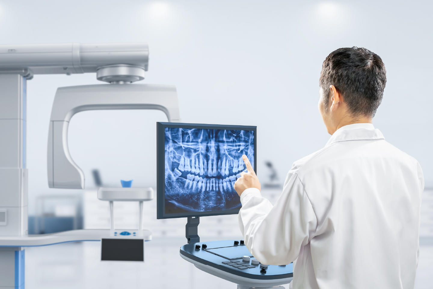
X-rays are an essential diagnostic tool in dentistry, allowing dentists to see areas of the teeth, gums, and jaw that are not visible during a standard oral examination. Digital X-rays are the modern approach to dental imaging, offering detailed images with reduced radiation exposure compared to traditional film X-rays.
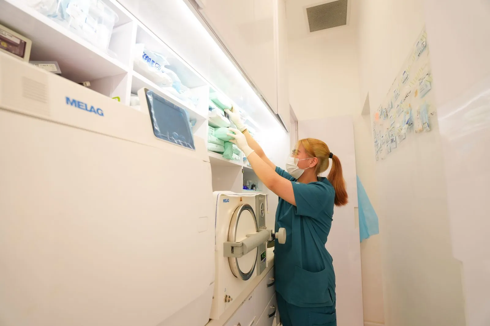
At Alpha Dental Group, digital X-rays are used to support accurate diagnosis, treatment planning, and preventive care. They help detect dental issues early, which can prevent more extensive procedures and preserve long-term oral health.
Digital X-rays use electronic sensors to capture images of the teeth, gums, and underlying bone structures. These images are instantly available on a computer screen, providing dentists with high-resolution detail that aids in precise diagnosis.
Unlike conventional film X-rays, digital X-rays require significantly lower levels of radiation, making them safer for patients while maintaining excellent image quality. They also allow for easy storage, sharing, and comparison over time, supporting ongoing monitoring of dental health.'
Bitewing X-rays are used to examine the upper and lower back teeth simultaneously. They are particularly helpful in detecting early signs of decay between teeth and assessing bone levels, which can indicate gum disease.
These X-rays provide a complete view of a single tooth, from the crown to the root tip and surrounding bone. Periapical images are valuable for diagnosing abscesses, infections, and root problems.
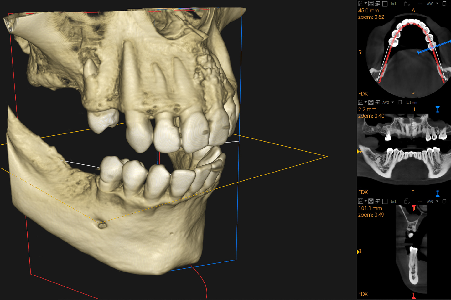
Panoramic X-rays capture a full view of the teeth, jawbones, and surrounding structures in a single image. They are useful for assessing tooth development, impacted teeth, jaw disorders, and planning certain dental procedures such as implants or orthodontics.
Primarily used in orthodontics, cephalometric X-rays show the relationship between the teeth, jaw, and skull. They assist in evaluating bite alignment and planning treatments that involve movement of teeth or jaw correction.
Digital X-rays allow dentists to identify cavities, fractures, infections, and bone loss that may not be visible during a clinical examination. Early detection makes treatment less invasive and helps preserve the natural teeth.
Digital imaging provides detailed information on bone density and structure, which is essential for diagnosing gum disease and planning procedures such as implants, extractions, or periodontal therapy.
Accurate imaging is crucial for treatments such as root canals, crowns, bridges, and orthodontics. Digital X-rays give dentists a clear view of tooth and jaw structures, enabling precise planning and predictable outcomes.
Digital X-rays require up to 80% less radiation than traditional film X-rays, making them a safer option for patients, including children and pregnant individuals, when necessary precautions are taken.
High-quality digital images can be viewed on a screen and explained to patients, helping them understand their dental condition and the rationale behind recommended treatments.
Before the procedure, a lead apron may be placed over the patient’s body to minimise radiation exposure. The dentist will position a small sensor or plate inside the mouth or use an external panoramic or cephalometric machine, depending on the type of X-ray required.
The digital sensor captures the image almost instantly, and the dentist can immediately review it on a computer screen. This allows for real-time evaluation and the opportunity to retake images if necessary.
Using the detailed images, the dentist can detect cavities, bone loss, infections, impacted teeth, or other abnormalities. The information supports informed treatment decisions and helps in monitoring changes over time.
Digital X-rays can be stored and compared with previous images to track the progress of treatments or the development of dental conditions, aiding in long-term oral health management.
While digital X-rays are safe, dentists will only recommend them when necessary. Routine dental examinations may not always require X-rays, and the frequency depends on age, dental history, and specific oral health needs. Pregnant patients should inform their dentist so that additional safety measures can be applied if imaging is required.
Digital X-rays are a vital part of modern dental care, offering a safe, efficient, and accurate method for diagnosing and monitoring oral health conditions. By providing detailed images with reduced radiation, they support early detection, precise treatment planning, and better patient understanding of dental conditions.
If you would like to learn more about digital X-rays and how they can support your dental health, contact Alpha Dental Group to arrange a consultation. Our dental team will assess your needs, explain the imaging process, and use the results to guide you toward the most suitable preventive or restorative care.